
Shenzhen Bineogen Technology Co., Ltd.
Bioimaging and data analysis solutions

Recent News
Heavy Research Express | Decrypt the neural circuit of chronic pain, Shenzhen Bineogen Lychnis software assists top breakthroughs!
Chronic pain is a major global public health challenge. Recently, top international academic journals published a breakthrough study. Scientists have completely resolved for the first time the driven chronic mechanical pain's "spinal cord-brain-spinal cord" multi-synaptic neural circuit. Findings about chronic pain's "exclusive circuit" why do wounds heal...
"Guangming News" Bineogen: Microscopic Imaging Technology Solves Brain Science Research Pain Points
Source: www.baoanone.com. Bineogen is dedicated to providing efficient, precise, and intelligent imaging solutions for researchers worldwide. Nowadays, global brain science is developing rapidly. Global brain science research is moving towards "mesoscopic scale whole-brain analysis...
Peking University National Biomedical Imaging Science Center's newly installed VISoR system has been debugged and put into use
Source: Peking University National Biomedical Imaging Science Center's VISoR system, based on light sheet scanning and synchronous real-time reading for efficient volumetric imaging, designed for high-resolution imaging of large biological samples, by Prof. Guoqiang Bi's research group, University of Science and Technology of China...
The world's fastest "high-definition CT" for the whole body of mice! Single nerve fibers are clearly visible
Professors Guoqiang Bi and Beiming Liu from the University of Science and Technology of China, Hefei National Research Center for Microscale Material Science and the School of Life Sciences and Medicine, in collaboration with the team from the Hefei Comprehensive National Science Center's Artificial Intelligence Research Institute and the Shenzhen Institutes of Advanced Technology, Chinese Academy of Sciences, have achieved significant breakthroughs in the field of large-scale biological tissue 3D microscopic imaging. The team has successfully developed
Training Review | 2025 Neural Circuit Tracing and Regulation Conference · Training Class
Shenzhen Bineogen Technology Co., Ltd. Training Course Introduction: The "2025 Neural Circuit Tracing and Regulation Conference · Training Course and Frontier Technology Innovation Conference on Acupuncture Research" successfully concluded in Shenzhen on May 30. This grand event focuses on cutting-edge technologies in the field of neuroscience, attracting many active researchers in neural circuit structure analysis...
Annotation and Visualization of 3D Whole Brain Imaging Data
Lychnis-viewer is a free software specifically designed for large-scale 3D/4D image data, mainly used for annotation and visualization of biological images such as neuron tracing. It supports multi-resolution data display and can efficiently process TB-level Imaris IMS documents...
probably in that order.”

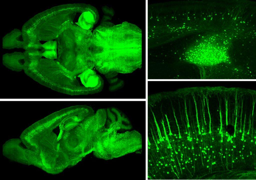
Whole brain clearing
After the improved clearing based on the CLARITY method, light passes through the brain slices without emission refraction, allowing uniform and clear 3D imaging within a thickness range of 300μm, revealing the fine structures inside the brain slices.

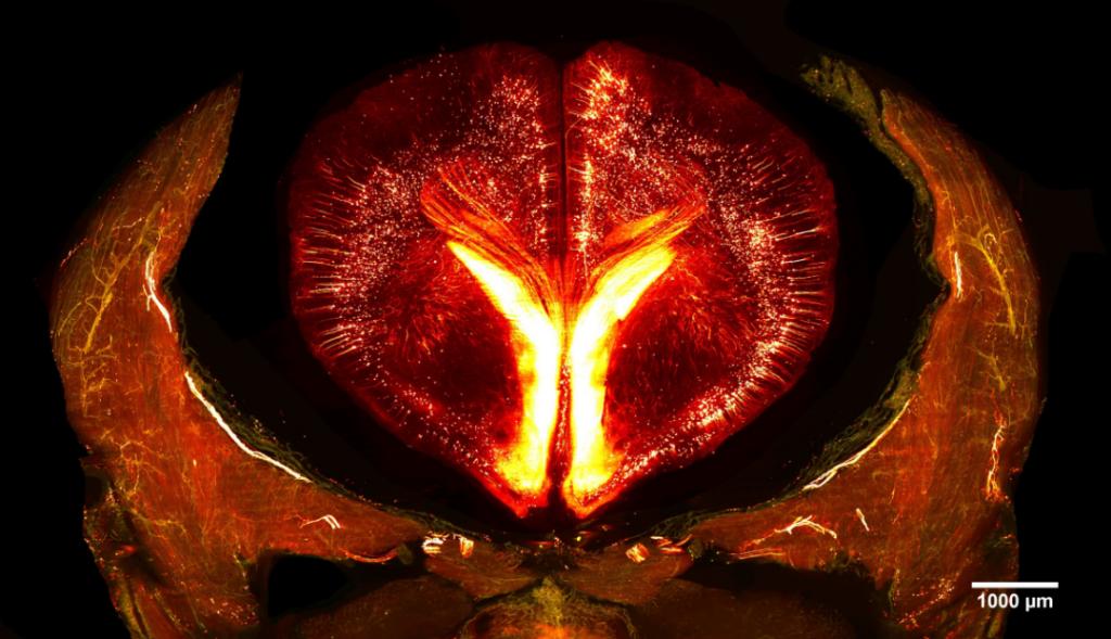
VISoR high-throughput 3D imaging
Using the VISoR high-throughput technology developed by Prof. Bi's team, we can complete whole mouse brain imaging at 1x1x2.5 μm3 resolution in just 0.5 hours.

Large-Scale Image Analysis
Using high-performance clusters and custom algorithms, we achieve storage, management, and analysis of large image data, including stitching, 3D reconstruction, registration, and neuronal reconstruction.
