Core technology
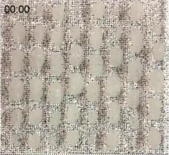
1. PuClear clearing Technology
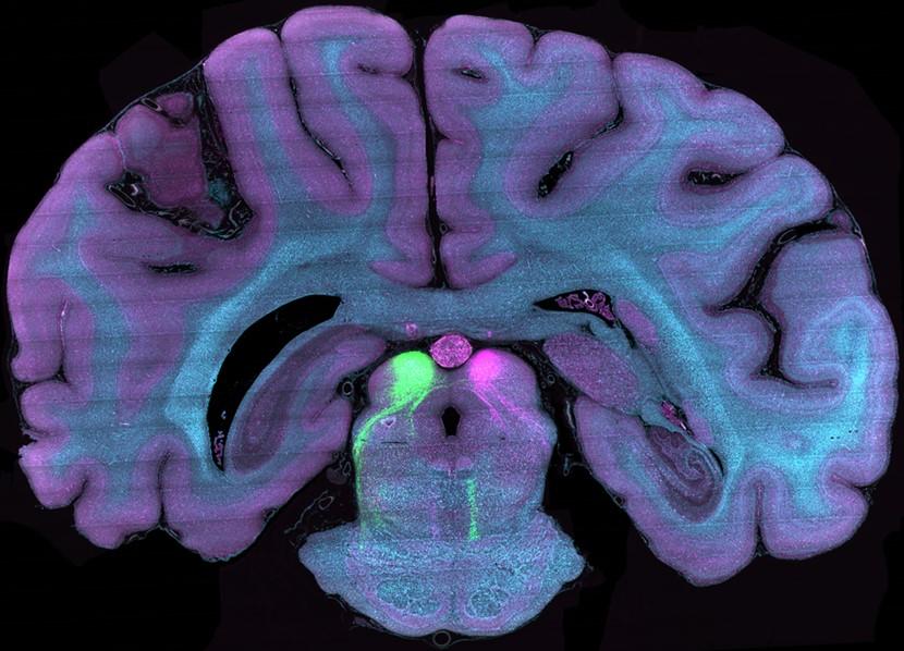
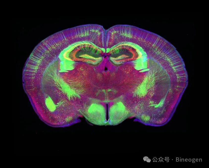
PuClear tissue clearing method is an improved clearing method based on hydrogel embedding above the CLARITY method. After postfixation and embedding, the proteins, nucleic acids, and other structures within the brain slices can be covalently bonded to the hydrogel framework, remaining in situ during subsequent clearing. After PuClear clearing, light does not refract when passing through the brain slices, allowing for uniform and clear 3D imaging within a thickness range of 300μm, and distinguishing the fine structures inside the brain slices.
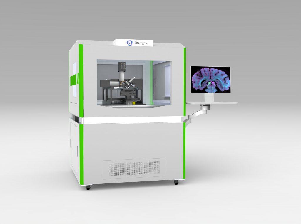
2. VISoR high-throughput 3D imaging
Using the VISoR high-throughput technology developed by Prof. Bi's team, we can complete whole mouse brain imaging at 1x1x2.5 μm resolution in just 0.5 hours.
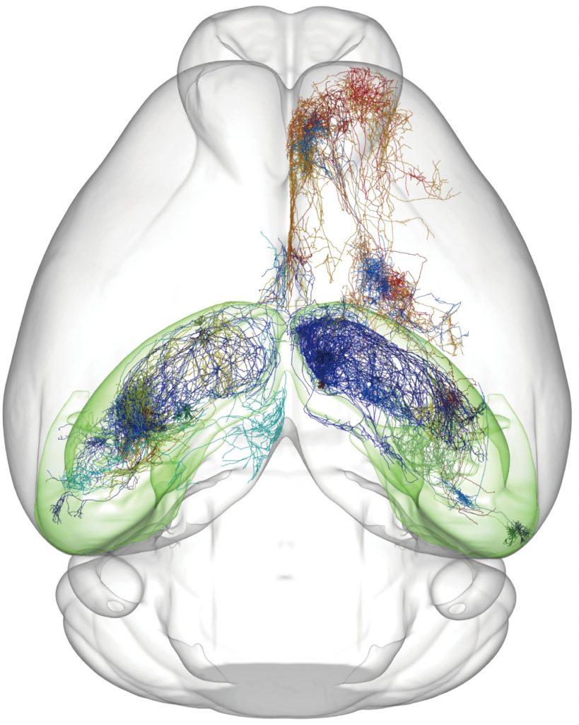
3. Large-Scale Image Analysis
We use the high-performance computing cluster we built, as well as self-developed algorithms and software, to achieve storage, management, and various analyses of large image data, including image stitching, 3D reconstruction, atlas registration, and neuron morphology reconstruction.
Highlight functions
PuClear clearing technology
- Covalently fixed structure:PuClear clearing technology uses covalent bonding to fix structures, binding proteins, nucleic acids, and other structures within the brain slices to the hydrogel framework.
- In situ structure preservation:During the clearing with PuClear technology, the structures within the brain slices are maintained in situ,
- Optical performance improvement:After PuClear clearing, light does not refract when passing through the tissue, allowing for clear and uniform 3D imaging. In contrast, traditional clearing methods may cause scattering or refraction of light, affecting imaging quality.
- Imaging range expansion: PuClear technology can process thicker tissue samples (such as brain slices), expanding the imaging range to a thickness of 300μm. Traditional clearing methods may not work well for thicker tissue samples, limiting the imaging range.

VISoR high-throughput 3D imaging
Multi-dimensional synchronized flying scan fluorescence microscopic imaging (VISoR) technology is a new type of rapid 3D micro-resolution imaging technology. It combines brain tissue clearing, synchronized imaging, and 3D reconstruction technology, capable of completing the analysis at the synapse level of the whole mouse brain in a short time with high resolution. The main features of this technology include:
- Synchronization scanning achieves continuous and clear imaging:Continuous and clear imaging was achieved during sample movement through synchronous scanning, greatly increasing the imaging speed to hundreds of times that of traditional imaging technologies.
- Fast imaging speed:Complete whole mouse brain imaging in 0.5 hours, with a voxel resolution of 1×1×2.5μm³.
- Multi-channel imaging:Applicable to samples of different sizes, allowing for multi-channel imaging, including tissue samples like mouse brains, with broad application prospects.
- Compatible with various labeling methods and can be extended to ultra-large samples:This technology is compatible with various labeling methods, applicable to different types of samples, and can even be extended to imaging of ultra-large samples.
Large-Scale Image Analysis
We use the high-performance computing cluster we built, as well as self-developed algorithms and software, to achieve storage, management, and various analyses of large image data, including image stitching, 3D reconstruction, atlas registration, and neuron morphology reconstruction. The image analysis system we built has the following characteristics:
- Processing large-scale:The underlying system uses a multi-level data storage structure to improve data access efficiency and processing speed, while reducing memory consumption for data processing.
- Large capacity storage:Data center with PB-level storage capacity, paired with high-performance CPU/GPU servers, can process massive amounts of data simultaneously.
- One-stop analysis platform:Includes a series of functional modules from image stitching, segmentation, atlas registration to video rendering, from data analysis to making article charts, without the need to purchase additional commercial software.