Source:Peking University National Biomedical Imaging Science Center
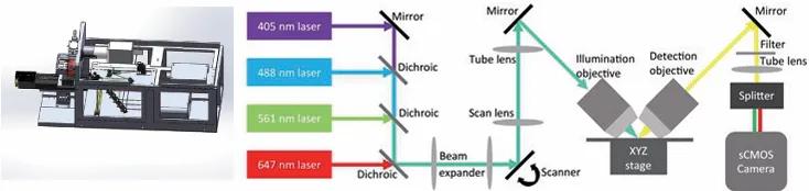
The synchronized flying scan 3D fluorescence microscopic imaging system (VISoR) is an efficient volumetric imaging system based on light sheet scanning and synchronized real-time reading, specially designed for high-resolution imaging of large-volume biological samples, developed by Professor Guoqiang Bi's research group at the University of Science and Technology of China. This device mainly achieves high-precision and rapid fluorescence imaging analysis of the internal structures of biological tissues and clinical medical samples such as brains, embryos, and pathological sections, and can perform high-throughput 3D fluorescence imaging of large samples. The VISoR imaging system has been installed and debugged in the public experimental platform on the first floor of Building 2 of the imaging facility and has officially started to provide technical services to the outside world. The main configuration and functions of the system are as follows:
Main functions and characteristics:
Light sheet microscopy system:Using a specific angle laser beam to excite the sample from the side to emit fluorescence, providing high-speed CMOS imaging, with the incident illumination optical path and CMOS fluorescence receiving optical path perpendicular to each other, achieving continuous and rapid imaging through the galvanometer, adjusting the optical path, and the three-dimensional displacement stage.
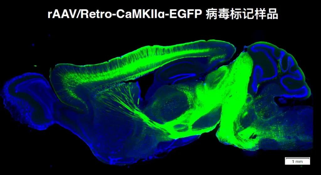
Whole brain fluorescence imaging:The mouse whole-brain fluorescence imaging service provides high-resolution, brain-wide imaging solutions for neural circuit research, neuron localization analysis, and neurological disease models. Utilizing VISoR high-throughput imaging and clearing technologies, we can obtain highly accurate three-dimensional imaging data, revealing the distribution of cells, neuronal connections, and distribution patterns throughout the entire brain.
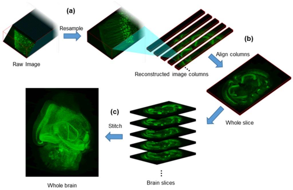
3D data processing:Using specially developed algorithmic techniques, the massive 2D data acquired was reconstructed into 3D, ultimately obtaining the 3D image of the sample.
Main specifications and technical indicators:
Resolution:Up to 0.5×0.5×2.5 µm³, ensuring clear imaging of microstructures;
Stage Travel:100 mm×150 mm, imaging range: 70 mm*75 mm;
Imaging System:Dual sCMOS cameras, 2048×2048 pixels, 100 fps full-frame imaging speed;
Imaging flux:Under high-resolution conditions, up to 1.0 T pixel data can be obtained every hour;
Imaging speed:Whole mouse brain imaging takes no more than 2 hours, greatly improving experimental efficiency;
Laser light source:Equipped with four wavelengths of lasers: 405 nm, 488 nm, 561 nm, and 640 nm, suitable for multi-channel fluorescence imaging;
Data acquisition workstation:Includes 32 TB SSD high-speed storage and 40 TB local storage.
Main application examples:
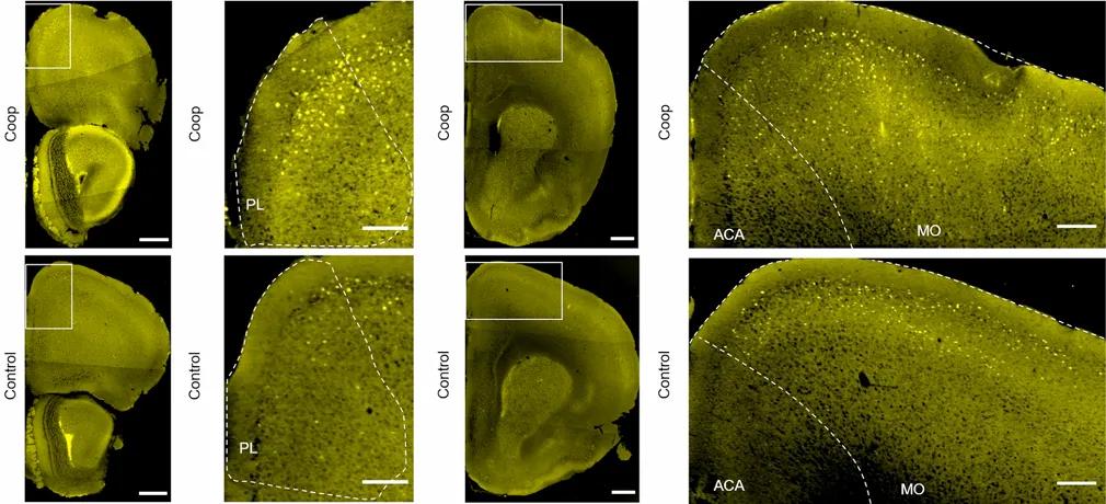
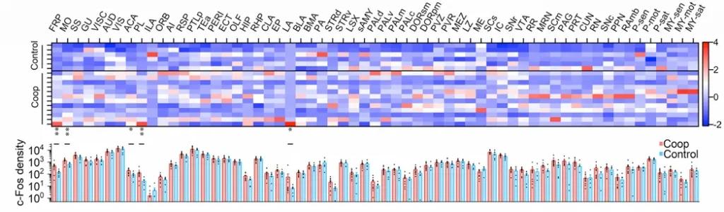
Brain activity patterns of mice in cooperative behavior:Using c-Fos labeling technology, we traced and analyzed the neural activity across the whole brain of mice in the cooperation and control groups, revealing the neural mechanisms behind the cooperative behavior.
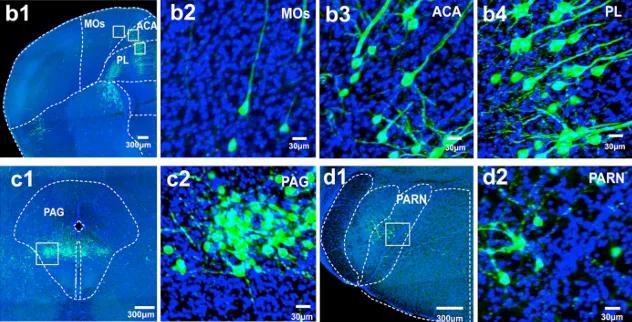
Whole brain VISoR imaging technology and retrograde tracing virus labeling:Revealing the whole-brain input distribution and axon branching patterns of layer 5 pyramidal neurons related to the mouse pedunculopontine nucleus (PPN), focusing on the projection characteristics of PPN-projecting neurons from the cortex to subcortical target areas, provides a unique perspective for understanding the functional connectivity between brain regions.
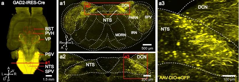
Remote projections of GABAergic neurons in the solitary tract nucleus:This study combines cell-type-specific viral tracing with VISoR high-resolution 3D imaging, and for the first time discovers that GABAergic neurons in the NTS project not only to the brainstem but also have long-range projection capabilities, targeting the bed nucleus of the stria terminalis (BST) and the paraventricular hypothalamus (PVH), and are involved in stress response and emotional regulation. Retrograde tracing and single-cell reconstruction further validate the origins of these long-range projections, providing new insights into the brainstem-forebrain pathways.
Technical references:
All image materials in this article are provided by Prof. Guoqiang Bi's research group.
1. Zhang, K.-M. et al. A new paradigm of learned cooperation reveals extensive social coordination and specific cortical activation in mice. Molecular Brain 16, 40 (2023).
2. Liu, Q.-Q. et al. Preferential subcortical collateral projections of pedunculopontine nucleus targeting cortical pyramidal neurons revealed by brain-wide single fiber tracing. Mol Brain 15, 88 (2022).
3. Shi, M.-Y. et al. Long-range GABAergic projections from the nucleus of the solitary tract. Mol Brain 14, 38 (2021).
Device Storage Location:
Peking University National Biomedical Imaging Science Center